
|
|
|||||
|
|
|||||
|
|
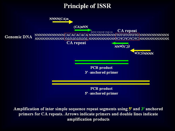
Analysis of Inter Simple Sequence Repeats (ISSR) PRINCIPLE The simple sequence repeats or microsatellites especially (TAA)n and (AT)n repeats are ubiquitous in the silkworm genome. The changes in the number of repeats in a microsatellite array, which is considered to be the result of slippage of the DNA polymerase which occurs during DNA replication, provide an unlimited source of polymorphism. However, studying SSR is quite labor intensive since complete sequence information flanking the repeats is necessary to design primers for PCR amplification. Here, we demonstrate an alternative, efficient method that does not require any prior sequence information. In this method microsatellite sequence anchored either at 5' or 3' ends with a stretch of degenerate nucleotides were used for inter-repeat region amplification.
ADVANTAGES
LIMITATIONS
Inter Simple Sequence Repeat region can be amplified using different protocols as per the lab’s requirement. In the past years we have tested many different protocols and standardised them for our population studies, polymorphism estimations, strain/variety identification, Mapping etc. Here, we list the best of our methods, which are tried and tested, in our lab for your use.
 I.Protocol of ISSR PCR for agarose gel electrophoresis 1. Equipment and reagents a) Thermal cycler b) Poly Acrylamide Gel Electrophoresis system c) The reagents including Taq DNA polymerase, 10 x PCR buffer (500 mM KCl, 100 mM Tris-HCl, 0.01% gelatin and 1% Triton X-100), 10 x dNTPs stock (1 mM) and 10 ng DNA samples. d) 1 x TBE (90 mM Tris borate, pH 8.3, 2 mM EDTA) e) Silver staining reagents like, fixative-stop solution (10% ethanol), silver nitrate solution (0.2% silver nitrate in water), developer solution (1.5% sodium hydroxide and 3ml/l formaldehyde). 2. ISSR PCR reaction mix
3. Thermal Cycling Conditions
Aliquots of amplified DNA from individual
PCR reactions should be loaded on a denaturing 2.5% (3 parts of Metaphor agarose: 1 part of agarose) gel in 1x
TBE. Electrophoretic separations should be performed in 1x TBE in a horizontal
gel tank. II. Protocol of ISSR PCR for PAGE and silver staining 1. Equipment and reagents a) Thermal cycler b) Poly Acrylamide Gel Electrophoresis system c) The reagents including Taq DNA polymerase, 10 x PCR buffer (500 mM KCl, 100 mM Tris-HCl, 0.01% gelatin and 1% Triton X-100), 10 x dNTPs stock (1 mM) and 10 ng DNA samples. e) Silver staining reagents like, fixative-stop solution (10% ethanol), silver nitrate solution (0.2% silver nitrate in water), developer solution (1.5% sodium hydroxide and 3ml/l formaldehyde). 2. ISSR PCR reaction mix
3. Thermal Cycling Conditions
Aliquots of amplified DNA from individual
PCR reactions should be loaded on a denaturing (7M Urea) 6% Poly Acrylamide
sequencing Gel in 1x TBE. After the bromophenol blue dye runs out, the gel
is silver stained for band detection.
5. Silver staining After electrophoresis, gels should
be fixed in fixative-stop solution for 30 min. Fixed gel must be rinsed 3
times with water for 2 min. each. Later gel must be impregnated with silver
nitrate solution for 10 min. and rinsed with distilled water for 5-20 sec.
Gel can be developed with cold developer solution for 4 min. Developing reaction
should be stopped with fixative-stop solution for at least 1 min. and washed
extensively with water. Dry stained gels at room temperature and store in
photographic albums. III. Protocol of ISSR PCR using radiolabel 1. Equipment and reagents a) Thermal cycler b) Long gel Poly Acrylamide Gel Electrophoresis (manual sequencing) system c) The reagents including Taq DNA polymerase, 10 x PCR buffer (500 mM KCl, 100 mM Tris-HCl, 0.01% gelatin and 1% Triton X-100), 10 dNTPs stock (1 mM) and 10 ng DNA samples. d) 1 x TBE (90 mM Tris borate, pH 8.3, 2 mM EDTA). e) Radiolabelled dATP
2. SSR PCR reaction mix
3. Thermal Cycling Conditions
Aliquots of amplified DNA from individual
PCR reactions should be mixed with formamide stop solution. Four micro-litre
of the sample must be denatured at 750C for 2 min., and immediately
chilled on ice. Electrophoretic separation must be done on 6% polyacrylamide
gel containing 8 M urea in 1 x TBE buffer. After electrophoresis, the gels
should be fixed for 2 x 20 min. with 10% glacial acetic acid. The fixed gel
must be air-dried and exposed for 4 -12 hrs. IV. Protocol for Fluorescent ISSR (FISSR) PCR** 1. Equipment and reagents a) Thermal cycler b) Automated sequencing System (e.g. ABI 377) c) The reagents including Taq DNA polymerase (preferably 'AampliTaq Gold' to prevent stutter bands, see Note below), 10 x PCR buffer (500 mM KCl, 100 mM Tris-HCl, and 1% Triton X-100), 10 x each of dGTP, dCTP dTTP and dATP stock (1 mM), 2m M stock of fluorescent dUTP (TAMARA, R110 or R6G, Perkin Elmer) and 5 ng DNA samples. d) 6 x loading buffer and GENESCAN-1000 ROX- labelled molecular weight standard e) 1 x TBE (90 mM Tris borate, pH 8.3, 2 mM EDTA)
3. Thermal Cycling Conditions
4. Sample preparation and electrophoresis conditions One micro-litre of PCR product should
be mixed with 1.5 m l of 6 X loading buffer (1:
4 mixture of loading buffer and formamide; Sigma). To this add 0.4 m l of GENESCAN-1000 ROX- labelled molecular weight
standard (red fluorescence). Before loading onto an ABI 377 automated sequencer,
the samples should be denatured at 92° C for
1 min. For an optimum separation, denaturing Polyacrylamide gel of 5% containing
7M urea in 1 X TBE buffer must be used. *NOTE 1: This is a sample reaction condition. Annealing
temperature and MgCl2 concentration varies for each primer set. |
||||||||||||||||||||||||||||||||||||||||||||||||||||||||||||||||||||||||||||||||||||||||||||||||||||||||||||||||||||||||||||||||||
| Copyright © 2004 All Rights Reserved, CDFD, Hyderabad, India | |||||||||||||||||||||||||||||||||||||||||||||||||||||||||||||||||||||||||||||||||||||||||||||||||||||||||||||||||||||||||||||||||||
 35 cycles